|
Phase contrast microscopes were created in 1934 by Fritz Zernike, a Dutch Physicist. Zeiss Optical Works was the first to manufacture these microscopes, during WWII. Before this invention, the brightfeild microscope was the one most used, but this microscope was used for fixed, stained specimens. The new phase contrast microscopes can be used for the viewing of live cells, at a higher level of structural detail viewed. This means that cell division can be viewed since the cells can be live, and high refractive index areas on the cell such as membranes and nuclei can be viewed more clearly. 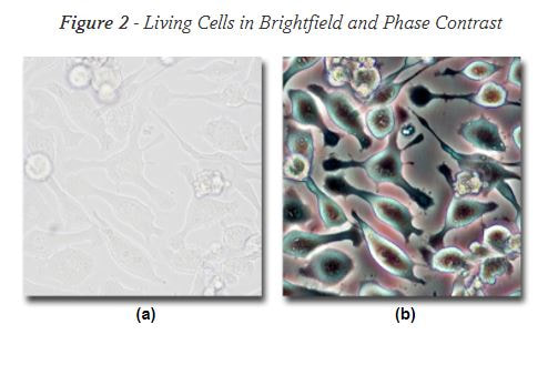 This kind of microscope produces a high contrast image. In general, as direct light passes through a specimen a phase change occurs. This phase change is converted into a change in brightness that allows specimens to be seen, even transparent ones. 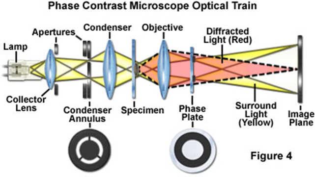
The diffracted light that is slowed by a quarter wavelength, and the surrounding light that is sped up by a quarter wavelength both assist in the contrast between the background and the specimen.
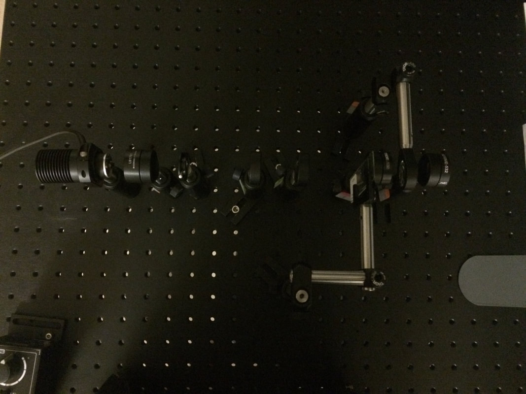
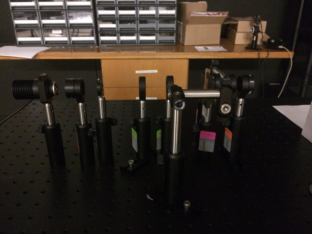
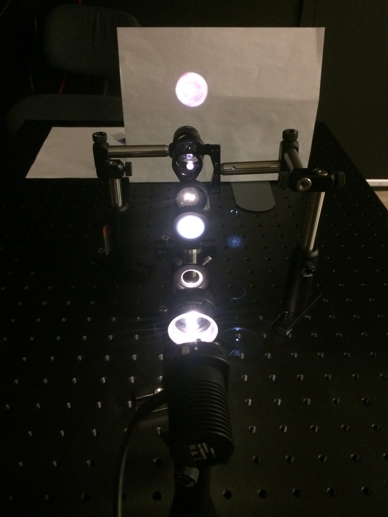 Through building this microscope I learned the importance of paying attention to focal lengths for the positioning of lenses. This was especially important for the first two lenses and their positions. Another realization that I had was the fact that the "objective lens" is actually comprised of two lenses that are very close together to produce the most clear image. For the largest, most clear image, the iris is opened enough so the light that reaches the condenser annulus is larger in diameter than the specially designed holes for light to go through. 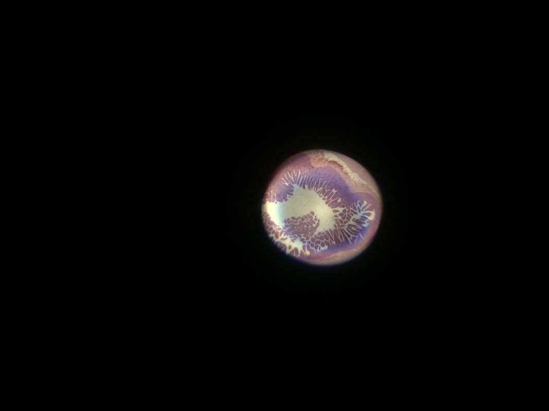 References:
0 Comments
|